Skin cancers are a rapidly growing portion of all cancers diagnosed each year. It is believed that increased sun exposure and a decreased ozone layer are a major cause of this phenomenon. This guide is designed to show you the common “warning” signs of skin cancer and to show pictures of cancer and pre-cancerous skin abnormalities. It is also designed to teach you how to avoid these cancers. The beautiful thing about skin cancer is that most are preventable, and those diagnosed early are usually curable. Unfortunately, those cancers not diagnosed early (especially melanoma) may be fatal! The key to skin cancer control is prevention, followed by early diagnosis.
By far the most common skin cancers are Basal Cell Carcinoma (BCC) and Squamous Cell Carcinoma (SCC). These two forms of skin cancer, along with malignant melanoma, make up the three skin cancers that afflict nearly a million Americans each year. Though basal cell carcinoma and squamous cell carcinoma are rarely fatal, they can cause a lot of damage to the skin and often can lead to disfigurement and the need for surgery. Metastatic melanoma is one of the most aggressive and deadly cancers around. The only way to cure it is to either prevent it in the first place, or have the early cancer surgically removed. Once the cancer has spread to other areas of the body (metastasized), it is much more difficult to cure.
Please consider filling out our anonymous survey once you have completed this guide. Keep in mind that this guide is NOT a substitute for seeing your physician if you have a skin or health problem about which you’re concerned. Because skin cancers are potentially deadly, there is no substitute for letting your doctor check out your skin on a regular basis. An undiagnosed or unrecognized skin cancer could kill you! One good rule-of-thumb is any growth or rash that has not healed after 3 weeks, or any changing or growing mole rash, should be evaluated by a doctor.
BASAL CELL CARCINOMA
How do I avoid getting it?
Prevention is the best way to avoid this disease. Though Basal Cell Carcinoma can develop anywhere on the body, and are not always caused by sun-damage to skin, the vast majority of new cases are caused by repetitive sun-exposure and sun-damage. Minimizing sun exposure, especially during the summer months, will significantly decrease your risk of triggering a Basal Cell Carcinoma. (Please refer to our page on minimizing sun exposure to learn exactly how to do this.) How’s it treated if I get it?
If you have abnormally appearing changes on your skin that you think might be an early Basal Cell Carcinoma you should contact your family physician. Suspicious appearing skin growths are often sampled (e.g., a small piece of skin is removed) and sent to the laboratory where a pathologist (a specialist at looking at diseases under the microscope) will look to see if there is any evidence of cancer. If there is, the treatment is destruction of the cancerous areas. This is usually done by surgically cutting out the diseased skin under local anesthesia. Occasionally, the cancerous skin will be destroyed by freezing it with liquid nitrogen, or by destroying it with electricity or a laser. For more extensive cancers, or for cancers involving cosmetically important areas (such as the nose, lip, or ear folds), microscopically-controlled surgery (Mohs’ Surgery) is often the best choice. Usually, BCC is cured once the cancerous skin is completely removed as this type of cancer rarely spreads (metastasizes) to other areas.
Additional Information For more information on Basal Cell Carcinoma, click here.
SQUAMOUS CELL CARCINOMA
What is it?
Squamous Cell Carcinoma (SCC) is the second most common skin cancer affecting over 200,000 people every year. In the United States, SCC usually develops in people over 55 years old (in Australia people in their 20s and 30s commonly get it). It is more common in men than women, and more common in fair-skinned than darker-pigmented folk. SCC is caused by exposure to carcinogens (cancer-causing agents. The most common carcinogen is sunlight. It is believed that SCC is related to the accumulated lifetime dose of solar radiation. The more sun you are exposed to, the more likely it is you’ll develop this skin cancer. Other carcinogens that cause SCC are arsenic, certain types of Human Papillomavirus (see our STD online Guide), certain types of tar, pitch, and fuel oils (used in industrial jobs), and other radiation exposure (x-rays and Gamma rays). Like all cancers, SCC is essentially normal skin cells that now are multiplying out-of-control. SCC commonly starts as a precancerous growth and slowly develops over time. It is most commonly found on sun-exposed areas of the body, especially the forehead, temple, ears, neck, shoulders, and legs (in those who have spent time sunbathing).
What’s it look like?
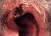
Squamous Cell Carcinoma usually begins as a precancerous growth and slowly develops into a firm small irritated lump. As this cancer grows, it commonly starts to breakdown and ulcerate. It will commonly bleed easily when scraped; it is usually painless, though may occasionally be tender or painful.
 |
 |
 |
|
Squamous Cell Carcinoma on the lower lip
|
Squamous Cell Carcinoma on the ear
|
Close-up of SCC on the arm
|
How do I avoid getting it?
Prevention is the best way to avoid this disease. Though Squamous Cell Carcinoma can develop anywhere on the body, and is not always caused by sun-damage to skin, the vast majority of new cases are caused by repeated sun-exposure and sun-damage. Minimizing sun exposure will significantly decrease your risk of getting Squamous Cell Carcinoma. (Please refer to our page on minimizing sun exposure to learn exactly how to do this.) Avoiding sunburns and wearing sunscreen on all exposed skin (with an SPF of 15 or better) is important to prevent getting this disease.
How’s it treated if I get it?
If you have abnormally appearing changes on your skin that you think might be an early Squamous Cell Carcinoma, you should contact your family physician. Suspicious appearing skin growths are often biopsied (e.g., a small piece of skin is removed) and sent to the laboratory where a pathologist looks to see if there is any evidence of cancer. If there is, the treatment is surgical removal of the cancerous tissue. Because SCC is more likely to spread if not removed early, it is important that the entire diseased area of skin be removed. Therefore, it is not recommended that SCC be frozen or destroyed as a tissue sample is important to be sure the whole thing was removed (e.g., “clean margins”). This is usually done by surgically cutting out the diseased skin under local anesthesia. Rarely, the cancerous skin will be destroyed by freezing it with liquid nitrogen. For more extensive cancers, or for cancers involving cosmetically important areas (such as the nose, lip, or ear folds) microscopically-controlled surgery (Mohs’ Surgery) is often used. This is done by a dermatologist or plastic surgeon specifically trained in this procedure.
Additional Information For more information on Squamous Cell Carcinoma, click here.
MALIGNANT MELANOMA
What is it?
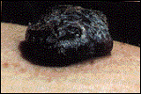
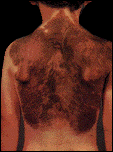
Malignant melanoma is the skin cancer that is most likely to metastasize (spread through the blood stream to other parts of the body). It is often triggered by repetitive sun exposure. With our societies preoccupation with tanning, malignant melanoma is unfortunately on the rise. (Some forms of melanoma are not sun-related; these forms of cancer have not been rising in incidence.) In addition to those exposed to sunlight, other people at risk for getting melanoma include people with a light complexion (freckles), light hair color with blue, green, or gray eyes, people with a history of a severe sunburn before age 20, a family history of melanoma or other skin cancer, or those with multiple, unusual, or congenital moles (a mole that some people are born with – see picture below).
What’s it look like?
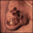
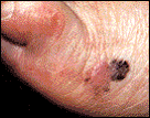
Melanoma takes many different forms. The most common is known as “superficial spreading” melanoma and usually begins as a tan spot that may slowly grow and change. Another type of melanoma is called “nodular” melanoma and usually develops in a black, blue or white mark that rapidly grows into a bump. To find out if your mole could be a melanoma, a doctor will need to examine you. If your mole is suspicious appearing (see The ABCDEs of melanoma to see what makes a mole suspicious), your doctor will likely take a small sample of it (biopsy) and/or refer you to a surgeon to do so.
 |
 |
 |
 |
|
Melanoma
(superficial spreading) of the nose |
Melanoma
(superficial spreading) of the foot |
Melanoma
(nodular) on the arm |
Congenital mole (nevus)
|
How do I avoid getting it?
Prevention is the best way to avoid this disease. Though malignant melanoma can develop anywhere on the body, and is not always caused by sun-damage to skin, the majority of new cases are caused by repetitive sun-exposure and chronic sun-damage. Minimizing sun exposure will significantly decrease your risk of getting melanoma. (Please refer to our page on minimizing sun exposure to learn exactly how to do this.) Avoiding sunburns and wearing sunscreen (SPF of 15 or better) on all exposed skin is important in preventing this disease. Keeping a good lookout for changing moles is the key to finding melanoma early.
How’s it treated if I get it?
Treatment of melanoma is initially surgical. Because melanoma can spread so quickly, EARLY DIAGNOSIS and removal of the suspicious or cancerous mole is crucial! (See The ABCs of finding suspicious moles.) Once melanoma has spread, it is much more difficult to treat. You should tell your doctor if you have any moles or new growths that are changing. Studies are underway to see if certain chemotherapies with or without radiation treatment can be effective, but thus far we do not have any great treatments for metastatic melanoma. Other research, including a vaccination that strengthens the body’s own response to the cancer, is currently underway.
Additional Information For more information on malignant melanoma, click here.
MOLE WARNING SIGNS
The “ABCD” rule and Melanoma Danger Signs
Finding melanoma early is the key to curing this vicious cancer. Learn the ABCD mnemonic for recognizing moles and growths that might be cancerous. Though most (if not all) of your “suspicious” moles will turn out to be normal non-cancerous moles, it is much better to be safe than to not see, or ignore, an early melanoma. Be sure to review how to do a monthly skin examination to properly look for abnormal growths.
If your mole or growth has one or more of the ABCDEs, you should show it to your physician as soon as possible!
Asymmetry
Asymmetry can be assessed by comparing one half of the growth to the other half to determine if the halves are equal in size. Unequal or asymmetric moles are suspicious.
 |
 |
|
Symmetric (normal)
|
Asymmetric
|
Border
If the mole’s border is irregular, notched, scalloped, or indistinct, it is more likely to be cancerous (or precancerous) and is thus suspicious.
 |
 |
|
Regular Border
(normal) |
Irregular Border
|
Color
Variation of color (e.g., more than one color or shade) within a mole is a suspicious finding. Different shades of browns, blues, reds, whites, and blacks are all concerning.
 |
 |
|
One Color (normal)
|
Color Variance
|
Diameter
Any mole that has a diameter larger than a pencil’s eraser in size (> 6 mm) should be considered suspicious.
Elevation
If a mole is elevated, or raised from of the skin, it should be considered suspicious.
Other Danger Signs of Malignant Melanoma
” Change in color, especially multiple shades of dark brown or black; red, white and blue,
” Change or spreading of color from the edge of the mole into surrounding skin.
” Change in size, especially sudden or continuous enlargement.
” Change in shape, especially development of irregular margins or border.
” Change in elevation, especially sudden elevation of a previously flat mole.
” Change in the surface texture of a mole, especially scaliness, erosion, oozing, crusting, ulceration, or bleeding.
” Change in the the surrounding skin, especially redness, swelling, or new moles.
” Change in sensation, especially itching, tenderness, or pain.
Basically, any mole or growth that is CHANGING needs to be checked by a doctor.
Examples
 |
This mole is Asymmetric and has an irregular Border. The middle part is Elevated off of the skin. It also has a Diameter larger than 6 mm. |
 |
This mole certainly is Asymmetric has an irregular Border. It has a larger Diameter than a pencil’s eraser. It has more than one shade of Color. |
 |
This growth has more than one Color and is thus suspicious. In addition, it is also Asymmetric and has an irregular Border. |
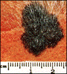 |
As you can see from the ruler, this mole’s Diameter is much larger than 6 mm (it measures 19 mm, or 1.9 cm in one direction, and probably is even larger if measured vertically). In addition, this mole is also Asymmetric and has a Border that is irregular. (Note, the presence or absence of hair in a mole is neither suspicious nor reassuring for cancer.) |
 |
This mole is obviously Elevated off of the skin. It’s diameter is also larger than a pencil’s eraser (6 mm). |
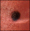 |
This mole is not elevated, it is symmetrical, of uniform color and has an essentially smooth border. It is smaller than 6 mm. It has none of the warning signs of melanoma, and is the only mole pictured that is “normal”. |
PRECANCEROUS GROWTHS
What is it?
There are a number of “precancerous” growths that, if left alone, may turn into skin cancer over time. Recognizing these precancerous skin growths and treating them will often avoid skin cancer altogether.
Dysplastic Nevus

One of the most concerning is the dysplastic nevus. These moles are quite common, and often run in families. They can be identified as they usually have one or more abnormal components (as described in The ABCDs section). (The dysplastic mole pictured has a border that is not smooth and has more than one color.) Dysplastic nevi have a propensity to turn into melanoma, though not all of them will. Yet given the seriousness of melanoma, it is recommended that dysplastic nevi be either removed or followed closely (e.g., have a doctor look at it every 3 – 6 months) to watch for any change that might indicate it is transforming into the deadly cancer.
Aktinic Keratosis
The most common precancerous skin growth is called an actinic keratosis (AK). Patients who have these commonly have a history of extensive cumulative sun exposure. The most common locations for these growths are the face, neck, rims of the ear, and on the backs of hands and forearms. The upper chest and upper back are also common sites. (Non-sunlight induced actinic keratoses can be caused by exposure to X-rays, radium, and other chemicals such as arsenic.)
What’s it look like?
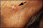
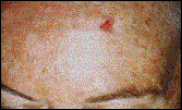
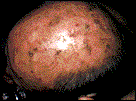
Actinic keratoses range in size from microscopic to several centimeters in size. They have a roughened sandpaper-like feel. They may be red, irritated, and mildly tender to touch.
 |
 |
 |
|
Actinic keratosis of forehead
|
Another AK of the forehead
|
Multiple AKs on the scalp
|
How do I avoid getting these?
Prevention is the best way to avoid these diseases. Though these growths can develop anywhere on the body, and are not always caused by sun-damage to the skin, many precancers are caused, or turn into cancer, by repetitive sun-exposure and chronic damage. Minimizing sun exposure will significantly decrease the risk of getting precancer and cancer. Avoiding sunburns and wearing sunscreen on all exposed skin (with an SPF of 15 or better) is important in preventing these diseases. Keeping a good lookout for changing moles is the key to finding melanoma early.
How’s it treated if I get it?
Treatment of dysplastic nevi is done by removing the abnormal mole or by close follow-up by the doctor. Removing dysplastic nevi is straight-forward, and can usually be done by your family physician in 15 minutes and with minimal discomfort. The procedure is as follows: the surrounding skin and atypical mole are cleaned and a small amount of lidocaine (Novocain, or similar) is injected shallowly into the skin. The mole is then gently cut out. A stitch or two may be needed depending on the size of the mole. Though it is called surgery, removing a small mole is as surgical as lightly skinning your knee (and actually hurts less).
Treatment of actinic keratosis is done by destroying the abnormal cells. This is usually done by freezing the skin with liquid nitrogen at the doctor’s office. Other techniques include using a prescription cream (5-flurouracil) on the abnormal skin. Interestingly, nearly 1 out of every 4 AKs will go away if the person simply avoids sun exposure.
Additional Information
For more information, click here.
PREVENTION

Each year nearly a million Americans develop skin cancer. Many more find that their skin is looking older, damaged, and wrinkled. All of these problems are preventable by avoiding the harmful effects of sun exposure. Now, this doesn’t mean you need to avoid the sun altogether – in fact, some direct sunlight is important in preventing other diseases, such as vitamin D deficiency. (Actually, you only need a few minutes of sunlight to get enough vitamin D, or you could even drink milk or eat tuna fish instead.) The problem is repetitive sun exposure. How many times did you go out on a sunny day last month? How about last year? What about over the last 5 years, 10 years? How many times did you get so much sun you got a sunburn? Though your tan (or sunburn) from last summer is long gone, the damage to your skin cells is still there. Unfortunately, repetitive sun exposure in the past adds up, and medical studies show that this repetitive exposure is a major risk for developing skin cancers.
How does the sun actually damage skin?
The harmful effects of the sun is from its ultraviolet (UV) radiation. When you expose your skin to the sun, your skin tries to protect itself (and you) by producing a chemical called melanin. This is a “natural” sunscreen that has a dark pigment that can reflect some of the dangerous ultraviolet light. Because the melanin pigment is dark, it makes your skin look dark, or tan. Those with blond hair or light skin types have less melanin than darker individuals and thus burn more easily – and have a much higher risk of skin damage and cancer. (Blacks have a lot of melanin, are therefore dark-skinned, and their risk of getting skin cancers are much less than Whites.) Unfortunately, melanin only partially protects the skin from the energy of the sun, and a “deep” tan doesn’t really add much protection. UV rays that are not reflected but absorbed by the skin can damage the DNA within the skin cells. Sometimes the damage is to a section of the DNA that tells the cell not to continue to grow and multiply (essentially an “off switch”). If this “off switch” for the DNA is broken, the cell literally keeps multiplying and growing. This is cancer.
How can I prevent myself from getting skin cancer?
Prevention is the key! The following three steps will help you avoid all of the skin cancers (and skin damage in general).
1. LIMIT YOUR EXPOSURE.
Try to limit or avoid the sun when its UV rays are the strongest (between 10 a.m. and 4 p.m.). A good rule-of-thumb is, if your shadow is shorter than you are, seek shade or cover up. Also, avoid tanning booths and sun lamps. UV light also goes through clouds, so just because it is cloudy doesn’t make you safe!
2. COVER UP
If you can’t avoid the sun, cover up your skin! Protect exposed skin by wearing clothes made of tightly woven, light-colored fabrics with long-sleeves and long pants. Wear a hat with a wide brim to protect your face and head. Wear sunglasses to protect your eyes and lower your risk of getting cataracts latter in life.
3. USE SPF-15 SUNSCREEN
For the parts of your skin not covered, use a waterproof sunscreen with a Sun Protection Factor (SPF) of at least 15. Don’t forget to use the sunscreen on your ears, lips, nose, and other areas that readily burn.

How do I watch for any suspicious moles?
Click here to see the 9 steps you should do monthly to find any early cancers or suspicious new growths.
SELF-SKIN EXAM
Review the explanation and diagrams below to learn the correct way to do a thorough skin examination. Print out our BODY DIAGRAM page and record any mole’s location. Each month when you recheck yourself, you can refer to your Body Diagram sheet to see if anything has changed. To begin, you should undress completely. Make sure the room is well-lighted and that you use a full-length mirror. A hand-held mirror and blow dryer will also be used. Any mole that is growing or changing should be shown to your family doctor. Click here for a list of melanoma warning signs (The ABCs).
 |
|
Step 1
|
Hold your hands with the palms up (see diagram). Look at your palms and between your fingers for any moles. Mark down any moles or discolorations on your Body Diagram sheet. Look at your forearms then turn over your arms and examine the backs of your hands, fingers, spaces between your fingers (webspace), fingernails and forearms.
 |
|
Step 2
|
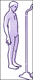
Stand in front of your full-length mirror. Hold up your arms, bent at the elbows, and view the backs of your forearms and elbows.
 |
|
Step 3
|
Using the full length mirror, observe the entire front of your body. Look at your face, neck, chest, groin, and legs.
 |
|
Step 4
|
Lift your arms over your head with the palms facing each other. Turn so that your right side is facing the mirror and look at the entire side of your body. Then turn and REPEAT on your left side.
 |
|
Step 5
|
With your back toward the mirror, look at your buttocks and the backs of your thighs and lower legs.
 |
|
Step 6
|
Using a hand-held mirror and the full-length mirror, view the back of your neck, your back, and your buttocks. Some areas are difficult to see, and it is helpful to have a partner assist you.
 |
|
Step 7
|
Using a hand-held mirror and the full-length mirror, view your scalp. Because it is difficult to view the scalp under your hair, use a blow-dryer (on cool setting) to lift the hair and get a better view of your skin below.
 |
|
Step 8
|
Sit down and prop your leg up on a stool or chair. Using the hand-held mirror, examine the inside of your leg and groin area, moving the mirror down the leg to your foot. REPEAT on your other leg.
 |
|
Step 9
|
Still sitting, cross one leg over the other. Use the hand-held mirror to examine the top of your foot, the toes, toenails and toe webspaces. Then look at the sole of your foot.
REPEAT on your other foot.
RESOURCES
This Skin Cancer Online Guide has allowed you to see photographs and learn about the most common skin cancers and precancers. The take home message is simple: Prevent yourself from getting skin cancer by being “sun smart”, and if you have a changing mole or non-healing scab, get it checked by a doctor ASAP (as soon as possible). All skin cancers are curable if caught before they spread. Once they have spread, treatment is much more difficult and usually includes surgery, chemotherapy and radiation treatment.
Some recommended readings on this topic can be ordered through Amazon.com.
1. Saving Your Skin: Prevention, Detection, & Treatment of Melanoma and Other Skin Cancer (by Barney Kenet, et. al.)
2. Cancer Free: The Comprehensive Cancer Prevention Program (by Sidney J. Winawer, M.D.)
For information on cancer in general, check out the following:
American Cancer Society
Thank you again for taking the time to check us out.
Please fill out the survey before you go.
BODY DIAGRAM
Print out the body template and use it to mark the location of your moles and skin abnormalities
 |
|
Do a self-skin exam every month
|
One Comment
Comments are closed.